EGI – Your EEG Research Partners
Welcome to EGI, where we have been at the forefront of EEG research and innovation for over three decades. With over 5,000 publications citing our EEG products, researchers choose EGI for a multitude of reasons. Our consultation method, researcher tools, brain-computer interface capabilities, and hyper-scanning options are just a few of the features that set us apart. Our innovative products allow researchers to go beyond traditional EEG measurements and delve deeper into understanding brain activity.
What makes our EEG Sensor Nets truly unique is their flexible design, catering to a wide range of subjects and patients. Equipped with elastomer forms, our sensor nets offer perfect fit and ease of use while providing accurate and reliable EEG measurements. Whether it’s quick recordings for infants with the rapid application sponged sensor nets, or capturing sleep EEG throughout the night with the classic gel application sensor nets, our high-density nets deliver exceptional results with maximum comfort.
At EGI, we understand the importance of integration in research. That’s why our EEG systems can seamlessly integrate with TMS, tDCS/tACS, fMRI and MEG systems, while also offering software tools for data analysis such as event-related potentials or electrical source imaging. Our sales and support team, equipped with a scientific background, are ready to offer advice and assistance to enhance your experimental setup. With a solution for every research budget and a range of EEG channel counts available (from 32-256), EGI is your partner in EEG research excellence.
Benefits of HD-EEG for Sensitive Groups, Patient Populations and Scientific Research
Geodesic Sensor Nets use abrasion-free application methods and the Net structure allows all electrodes to be applied at once. Application times for the routine (saline) HydroCel Geodesic Sensor Net range between 5 minutes for 32 channels to 10-15 minutes for 256 channels. EEG staff rapidly learn the application technique during on-site training when a HD-EEG system is purchased and additional training videos can be found on our website (egi.com).
Depending on the population you are working with, our knowledgeable team can provide recommendations tailored to your plans based on nearly 30 years of experience with sites around the world.
In study after study, patients and research participants report that they prefer EEGs performed with Geodesic Sensor Nets over conventional EEG sensors. People report that GSNs are more comfortable than other traditional clinical and research EEG methods.
- The Geodesic Sensor Net is a preferred option for infant EEG as the short application time and full head coverage allows infants to fall asleep which is suitable for many infant recordings. Saline is also gentler on sensitive infant skin compared to gel, reducing the risk of later discomfort after the session is complete.
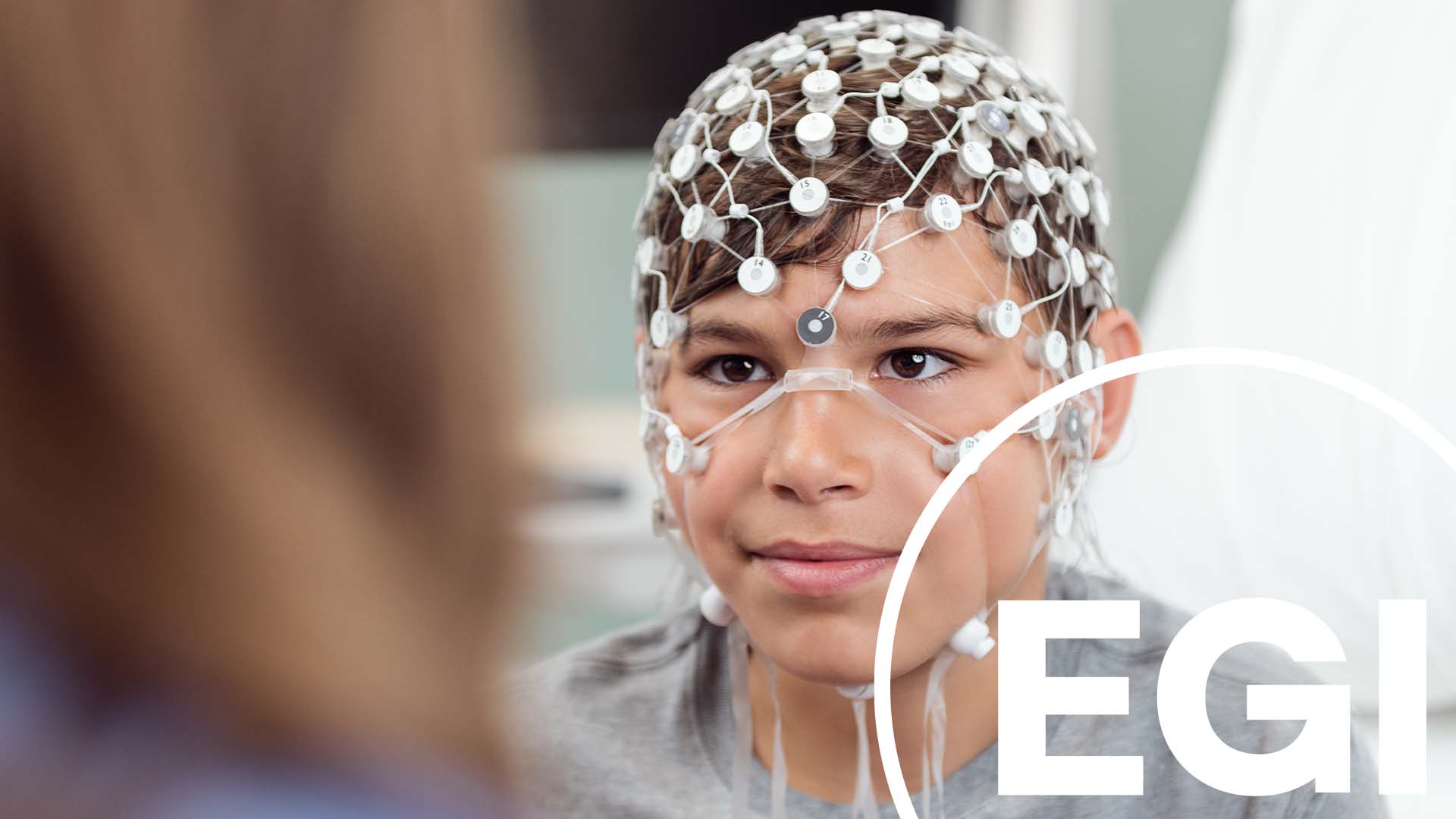
- Children are far less likely to cry or refuse the EEG with HD-EEG sessions using the Geodesic Sensor Net because the electrode application process is rapid (a few minutes), abrasion-free and requires less adjustment time before the recording. This fast, painless process also improves the likelihood that patients and research subjects will return for follow up visits.
- Geodesic Nets have been used successfully with difficult-to-record populations, such as children with autism, genetic disorders and sensory sensitivities, elderly patients with dementia, and people with ADHD.
- Patients also comment that the saline electrolyte used with routine EEG Nets does not smell or damage their hair. Hair washing is not required after saline-based HD-EEG sessions whereas it is required for traditional gel or paste based EEG.
Because no abrasion is required with Geodesic Sensor Nets, the skin on the scalp is not compromised, which reduces the risk of infection. Often, it is argued that mild abrasion during the course of skin preparation does not compromise the integrity of the skin. However, studies have shown that this is not an accurate assumption.
For further information, see Bild, S. (1997). Detection of occult blood on EEG surface electrodes. American Journal of Electroneurodiagnostic Technology, 38, 251-257.
Sedating children to obtain EEG is often done in clinical settings. This is not required when you use Geodesic Sensor Nets; rapid application time and the comfortable fit make sedation unnecessary.
The unique open and flexible design of the geodesic sensor net makes HD-EEG suitable for all ages and hair types. In 2024, EGI released a modifed “tall” version of the geodesic sensor net optimized to make better contact with the scalp for dense, curly and textured hair that might otherwise lift the electrode off the scalp, resulting in poor data quality. A 2023 study conducted by Northwestern University demonstrated the effectiveness of these new “tall” sensor nets in achieving lower impedance and better data quality.
Complete head coverage ensures that all clinically relevant info is captured. Guaranteeing complete head coverage requires attention to both appropriate intersensor distance and coverage of the underside of the head.
The first aspect of complete head coverage refers to the intersensor distance (that is, how far apart one sensor is from another sensor). Smaller intersensor distances translate to a more accurate measurement of the voltage field.
The second aspect refers to coverage of the underside of the head (for example, below the ears, including the face). It is common to believe that electrodes on the inferior surface of the head do not pick up EEG. However, voltage fields generated by the brain are volume conducted throughout the entire head. Moreover, certain brain regions, such as the inferior temporal lobes and ventral aspects of the frontal lobe, are oriented such that the voltage fields generated by these brain regions are best detected by electrodes on the inferior surface of the head.
The consequence of incomplete head coverage is that significant EEG signals can be missed by sparse electrode placement and by lack of electrode placement on the underside of the head. For detailed information, see:
Luu, P., Tucker, D. M., Englander, R., Lockfeld, A., Lutsep, H., & Oken, B. (2001). Localizing acute stroke-related EEG changes: Assessing the effects of spatial undersampling. Journal of Clinical Neurophysiology, 18, 302-317.
Sperli, F., Spinelli, L., Seeck, M., Kurian, M., Michel, C. M., & Lantz, G. (2006). EEG source imaging in pediatric epilepsy surgery: A new perspective in presurgical workup. Epilepsia, 47, 1-10.
Srinivasan, R., Tucker, D. M., & Murias, M. (1998). Estimating the spatial Nyquest of the human EEG. Behavior Research Methods, Instruments, & Computers, 30, 8-19.


